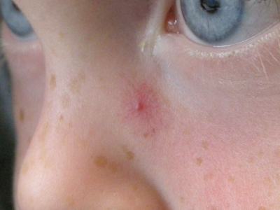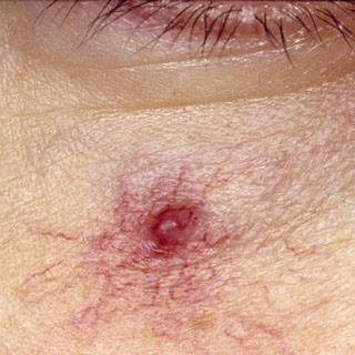
Spider naevi (also called spider angiomas) describe a central red papule with surrounding capillaries. The lesions blanch upon pressure. Spider naevi are almost always found on the upper part of the body.
Spider naevi can be differentiated from telangiectasia by pressing on them and watching them fill. Spider naevi fill from the centre, telangiectasia from the edge .
Around 10-15% of people will have one or more spider naevi and they are more common in childhood. Other associations